Table of Contents >> Show >> Hide
- Melanoma Diagnosis 101: The Big Picture
- Step 1: Spotting the Clues (and Knowing When to Get Checked)
- Step 2: The Clinical Evaluation (Your Story + A Close Look)
- Step 3: The Biopsy (The Only Way to Confirm Melanoma)
- Step 4: The Pathology Report (Where the Diagnosis Becomes Official)
- Step 5: Staging After Diagnosis (Do You Need More Tests?)
- Step 6: Practical PrepHow to Make Your Appointment More Productive
- Common Myths That Make Dermatologists Sigh (Gently)
- Real-World Experiences: What the Diagnosis Process Actually Feels Like (500+ Words)
- Conclusion: The Most Important Takeaways
Quick (but important) disclaimer: This article is for education, not medical advice. If you have a spot that worries you, the best next step is a clinicianideally a dermatologistwho can examine it and decide whether a biopsy is needed.
Melanoma is the “take it seriously” member of the skin-cancer family. The good news: when it’s found early, it’s often very treatable. The tricky part: melanoma can be sneaky. It can look like a harmless mole, a freckle that got ambitious, or a spot that seems to appear out of nowhere like it just moved into your skin without telling you.
So how is melanoma diagnosed? In one sentence: doctors diagnose melanoma by examining your skin and confirming suspicion with a biopsy that is reviewed under a microscope by a pathologist. Everything elsedermoscopes, photos, fancy imagingsupports that core process.
Melanoma Diagnosis 101: The Big Picture
Most melanoma diagnoses follow a pretty consistent arc:
- Someone notices a suspicious spot (you, a partner, a barber, a nail tech, your primary care clinicianshout-out to the people who save lives during regular life).
- A clinician evaluates it with a focused history and a skin exam (often with dermoscopy).
- A biopsy is performed to remove all or part of the lesion.
- A pathologist examines the tissue and produces a report that confirms melanoma or rules it out.
- If melanoma is confirmed, staging tests may follow depending on tumor features (thickness, ulceration, etc.) and your clinical situation.
That’s the overview. Now let’s zoom inbecause the details matter, and they can be the difference between “we’ll keep an eye on it” and “let’s take this off today.”
Step 1: Spotting the Clues (and Knowing When to Get Checked)
The ABCDE Rule (A Handy Checklist, Not a Final Verdict)
Clinicians often teach the ABCDE rule to help identify moles that deserve a closer look:
- A Asymmetry: one half doesn’t match the other.
- B Border: irregular, scalloped, notched, or blurry edges.
- C Color: multiple colors (brown/black with red, white, blue, or gray mixed in).
- D Diameter: often larger than about 6 mm (the size of a pencil eraser), but melanoma can be smaller.
- E Evolving: changing in size, shape, color, or symptoms (itching, bleeding, crusting).
Two key points people miss:
- “D” is not a free pass. Small lesions can still be melanoma.
- “E” is the drama queen of the group. Evolutionchange over timeis one of the most meaningful red flags.
The “Ugly Duckling” Sign
Another useful concept is the ugly duckling: a spot that looks different from your other moles. If most of your moles are similar and one is doing its own weird solo career, that’s worth checking.
When to Seek Care Promptly
Make an appointment sooner (not “sometime this year”) if you notice:
- a new, rapidly growing dark spot
- a mole that bleeds, oozes, crusts, or won’t heal
- an existing mole that changes noticeably over weeks to months
- a spot with multiple colors or a very irregular edge
- a pigmented streak under a nail that’s widening or changing
Step 2: The Clinical Evaluation (Your Story + A Close Look)
The History: The Questions That Actually Matter
Expect questions like:
- When did you first notice the spot?
- Has it changed (size, shape, color, symptoms)?
- Has it bled, itched, or become painful?
- Do you have a personal or family history of melanoma?
- Do you have many moles, atypical moles, or significant sun exposure/tanning-bed use?
- Are you immunosuppressed (for example, certain medications or organ transplant history)?
This isn’t small talkthese answers influence how suspicious a lesion is and how urgently it should be biopsied.
The Skin Exam: Full-Body, Not Just the “One Spot” You Came In For
A careful clinician often does a total body skin exam, because people can have more than one concerning lesion. They may also examine:
- the scalp (yes, they might part your hair)
- between fingers and toes
- soles of feet and palms
- nail beds
- behind ears and along the back
If melanoma is suspected or confirmed, the clinician may also check nearby lymph nodes (neck, armpits, groin) for enlargement.
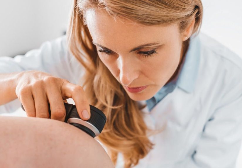
Dermoscopy: A Magnifying “Clue Finder”
Many dermatologists use a dermatoscope (dermoscopy) to see structures beneath the skin surface that the naked eye can’t. Dermoscopy doesn’t “diagnose” melanoma by itself, but it can significantly improve clinical evaluation and help decide whether a biopsy is needed.
In some settingsespecially for people with lots of molesclinics may also use photography (total-body photos) or digital mole monitoring to track change over time. Helpful? Yes. A replacement for biopsy when cancer is suspected? No.
Step 3: The Biopsy (The Only Way to Confirm Melanoma)
Here’s the blunt truth, delivered gently: you can’t confirm melanoma without a biopsy. That means removing tissue so it can be examined under a microscope. Even the most experienced dermatologist can’t (and shouldn’t) rely on vibes alone.
The Goal: Get Enough Tissue to Measure Depth
Why do clinicians care so much about biopsy technique? Because melanoma isn’t just about “is it cancer?”it’s also about how deep it goes. Depth (called Breslow thickness) is one of the most important features for staging and next-step decisions. If a biopsy is too shallow, it may miss the deepest portion and make staging less accurate.
Common Biopsy Types for Suspicious Pigmented Lesions
1) Excisional Biopsy (Often Preferred When Feasible)
An excisional biopsy removes the entire lesion with a small rim of normal-appearing skin. For many suspicious moles, this is the cleanest way to get a full specimen for the pathologist.
2) Deep Shave (Saucerization) Biopsy
A deep shave (sometimes called saucerization) removes a deeper “scoop” of skin than a superficial shave. In experienced hands, it can capture enough depth for accurate diagnosis and measurementespecially on areas where a traditional excision might be challenging.
3) Punch Biopsy
A punch biopsy uses a circular tool (think tiny cookie cutter, but medical) to remove a core of skin. It can be useful for very large lesions when removing the whole thing immediately isn’t practical. The downside: sampling error. If the punch misses the most suspicious area or the deepest focus, the pathology may underestimate the lesion.
4) Incisional Biopsy
An incisional biopsy removes only part of the lesion (with a scalpel). Like punch biopsies, it’s typically reserved for situations where full removal isn’t feasible right away.
What About a Superficial Shave Biopsy?
Superficial shaves are often great for many non-melanoma skin conditions. For a lesion strongly suspicious for melanoma, clinicians generally aim for a technique that captures full thickness so depth can be measured. If a shave technique is used, it needs to be deep enough to avoid missing the true thickness.
What the Biopsy Appointment Usually Feels Like
- Numbing: local anesthetic injection (brief sting, then numb).
- Removal: you may feel pressure, not pain.
- Closure: stitches for many excisions/punches; sometimes no stitches for a deep shave.
- Aftercare: keep it clean, apply ointment if advised, watch for infection signs.
Most biopsies are quick outpatient procedures. The waiting is usually the hardest part.
Step 4: The Pathology Report (Where the Diagnosis Becomes Official)
Once the specimen reaches the lab, a pathologist examines it under a microscope. If melanoma is present, the report often includes specific features that guide staging and treatment planning.
Key Terms You Might See
Melanoma in Situ vs. Invasive Melanoma
- Melanoma in situ: abnormal melanocytes are confined to the top layer of skin (epidermis). It hasn’t invaded deeper tissues.
- Invasive melanoma: cells have invaded into the dermis (deeper skin), which increases the risk of spread.
Breslow Thickness (Depth in Millimeters)
Breslow thickness measures how deep the melanoma extends into the skin. This measurement is central to staging decisions and can influence whether sentinel lymph node biopsy is discussed.
Ulceration
Ulceration refers to breakdown of the skin over the tumor and is considered a higher-risk feature in staging.
Margins
The report may comment on whether melanoma cells reach the edges (“margins”) of the specimen. Positive margins can mean additional surgery is needed to fully remove the cancer.
Mitotic Rate and Other Features
Depending on the case, reports can include details such as mitotic activity (how quickly cells are dividing), lymphovascular invasion, regression, or subtype (like superficial spreading melanoma, nodular melanoma, lentigo maligna melanoma, acral lentiginous melanoma).
Why Second Opinions Sometimes Happen
Melanocytic lesions can be complex. In borderline or unusual cases, clinicians sometimes request a dermatopathology second opinionespecially if the diagnosis impacts major decisions (wide excision margins, sentinel node biopsy, systemic therapy).
Step 5: Staging After Diagnosis (Do You Need More Tests?)
If biopsy confirms melanoma, the next question becomes: has it spread, and what stage is it? Early-stage melanoma may need only surgical management. Higher-risk cases may require additional staging steps.
Wide Local Excision (Often the Next Step)
After melanoma is diagnosed, many patients undergo a wide local excisiona procedure to remove additional normal-appearing skin around the melanoma site to reduce recurrence risk. The recommended margin depends on tumor characteristics and stage.
Sentinel Lymph Node Biopsy (SLNB)
A sentinel lymph node biopsy checks the first lymph node(s) that drain the melanoma areathe “sentinel” nodes. If melanoma has started to spread, these nodes are among the most likely early destinations.
SLNB isn’t for everyone. It’s typically discussed when risk of nodal spread is high enough to justify the procedureoften influenced by Breslow thickness and other high-risk features. Your team weighs the potential staging benefit against the fact that it’s a surgical procedure with its own risks.
Imaging and Lab Tests
For many early melanomas, routine scans aren’t necessary. For higher-stage disease or concerning symptoms, clinicians may order imaging such as:
- CT scans
- PET/CT
- MRI (especially if there are neurologic symptoms)
Some patients may also have blood tests (like LDH in specific contexts) as part of staging or monitoring, depending on clinical scenario.
Molecular Testing (When It’s Relevant)
If melanoma is advanced or has spread, doctors may test tumor tissue for mutations (such as BRAF) that can guide targeted therapy options. This isn’t always necessary for an early, thin melanoma, but it becomes important in certain stages and treatment planning.
Step 6: Practical PrepHow to Make Your Appointment More Productive
Bring These “Receipts” If You Have Them
- Photos showing how the spot changed over time (date-stamped if possible)
- A list of personal and family skin-cancer history
- Medication list (especially immune-suppressing meds)
- Notes on symptoms (itching, bleeding, tenderness, rapid growth)
Ask Smart Questions (Without Feeling Like You’re “Being Difficult”)
- What makes this lesion concerning (or not concerning)?
- Do you recommend biopsy? If yes, which type and why?
- When will results be back, and how will I get them?
- If it’s melanoma, what are the next steps (excision, SLNB discussion, referral)?
Reminder: advocating for clarity is not the same thing as being annoying. You’re allowed to understand your own skin.
Common Myths That Make Dermatologists Sigh (Gently)
Myth 1: “If it doesn’t hurt, it can’t be serious.”
Melanoma often doesn’t hurt. Pain is not a reliable safety signal.
Myth 2: “A blood test can tell me if my mole is melanoma.”
For a suspicious skin lesion, diagnosis is made by biopsy and pathology. Blood tests are not a stand-alone diagnostic tool for a new mole.
Myth 3: “My dermatologist can tell 100% without a biopsy.”
Even experts use tools (dermoscopy) and pattern recognition, but confirmation requires tissue under a microscope.
Myth 4: “If it’s melanoma, it will be huge and jet-black.”
Some melanomas are small, subtle, or even pink/red (amelanotic). That’s why “evolving” and “ugly duckling” matter.
Real-World Experiences: What the Diagnosis Process Actually Feels Like (500+ Words)
Medical flowcharts make melanoma diagnosis look tidy: exam → biopsy → report → next steps. Real life is messierand if you’re in it, you deserve to know that what you’re feeling is normal.
The “Is This Even Worth Asking About?” Moment
A lot of people don’t march into a clinic confidently announcing, “Greetings. I believe this is melanoma.” They hesitate. The spot is small. They’re busy. They Google. They un-Google. They ask a friend. The friend says, “Probably nothing,” which is the unofficial motto of procrastination.
One common story goes like this: a person notices a mole that looks slightly differentmaybe darker, maybe a new edge, maybe it started itching. But it’s not dramatic. It’s not painful. And they don’t want to be “that person” who makes an appointment for a freckle. Then a month goes by. Then three. Then the mole bleeds after a towel rub, and suddenly it’s very appointment-worthy.
If you recognize yourself in that timeline: you’re not alone. And you’re not wasting anyone’s time by getting checked.
The Appointment: Relief, Vulnerability, and a Weirdly Bright Room
Skin exams can feel surprisingly vulnerableespecially if you weren’t expecting a full-body check. Many people feel self-conscious about scars, body changes, or just the awkwardness of standing under bright lights while someone inspects your freckles like they’re reviewing a cosmic map. Clinicians do this all day; patients do not. It’s okay to say, “I’m a little nervous,” and it’s okay to ask what’s happening as it happens.
When dermoscopy comes out, some patients feel immediate reassurance (“They’re being thorough!”), while others feel the opposite (“Oh no, the fancy magnifier is herethis is serious.”). Both reactions are normal. Dermoscopy can simply mean your clinician is doing a higher-quality assessment, not that they’ve already decided the outcome.
The Biopsy: The Sting, the Pressure, and the Emotional Hangover
People often fear the biopsy procedure itself, but the physical part is usually brief. The numbing injection stingsno point sugarcoating itbut then the area gets numb quickly. Most patients describe the rest as pressure and tugging rather than pain. The bigger emotional hit often comes after, when you look at the bandage and think, “Okay… now we wait.”
Waiting for pathology results can feel like your brain has opened 37 tabs and none of them will load. Some people refresh patient portals like it’s a competitive sport. Others avoid looking because they’re afraid of what they’ll see. Sleep can be weird. Appetite can be weird. You might swing between “It’s probably fine” and “I am absolutely doomed” in the same afternoon. That doesn’t make you dramaticit makes you human.
When It’s Not Melanoma (Yes, That Happens a Lot)
Here’s a truth that rarely makes headlines: many biopsied lesions are benign. Clinicians would rather biopsy a suspicious spot and be wrong than ignore it and be tragically right. If your results come back benign, you may feel relief… and then irritation (at the scar, at the stress, at the cost, at the time). That’s also normal. You didn’t “overreact.” You acted responsibly with incomplete information.
When It Is Melanoma: A New Language You Learn Fast
If the biopsy confirms melanoma, many patients describe an immediate shift into “learning mode.” You suddenly hear words like Breslow thickness, ulceration, margins, and sentinel lymph node biopsy. You might feel overwhelmed, but you’ll also likely find that care teams are used to explaining this step-by-step. Writing down questions, bringing a support person, and asking for a copy of the pathology report can help you stay grounded.
Perhaps the most common reflection from people who’ve been through it: “I wish I’d come in sooner.” Not because earlier is always life-or-death, but because earlier usually means simplersmaller procedures, fewer decisions, less uncertainty. If you’re reading this with a suspicious spot in mind, consider that your future self may be very grateful you booked the appointment.
Conclusion: The Most Important Takeaways
Melanoma diagnosis isn’t magicit’s a process. It starts with noticing change, continues with a careful clinical exam (often aided by dermoscopy), and is confirmed by biopsy and pathology. If melanoma is found, the pathology details guide what happens next, from a wider excision to possible sentinel lymph node biopsy or imaging in higher-risk situations.
If you’re worried about a mole, don’t try to out-Google your skin. Get it examined. The best-case scenario is peace of mind. The worst-case scenario is exactly why early evaluation matters.