Table of Contents >> Show >> Hide
- First, a quick reality check: Is 16 weeks a “standard” ultrasound?
- Why you might have a 16-week ultrasound
- How to prepare (without overthinking it)
- What happens during the ultrasound appointment
- What you’ll see at 16 weeks
- What your provider is checking (the “why we’re here” list)
- What happens after the ultrasound
- Safety and limits: what ultrasound can (and can’t) do
- FAQ: quick answers to common 16-week ultrasound questions
- Real-life experiences at a 16-week ultrasound (about )
- Conclusion
You’re 16 weeks pregnant, your bump is finally making a statement, and now you’ve got an ultrasound on the calendar.
Cue the excitement… and the oddly specific question everyone ends up asking: “Do I need to pee before this or not?”
(Answer: it dependsmore on that in a minute.)
A 16-week ultrasound can be a sweet sneak peek into the second trimestersometimes scheduled as a follow-up,
sometimes done for a specific medical reason, and sometimes just because your provider wants an earlier look before
the big anatomy scan later on. Either way, it helps confirm how things are going, checks key measurements, and gives
you a chance to watch your baby do tiny, unbothered gymnastics on a screen.
First, a quick reality check: Is 16 weeks a “standard” ultrasound?
In the United States, many people have at least one ultrasound in pregnancy, and the most detailed routine scan is
commonly done in the second trimesteroften around 18–22 weeks. That’s the “anatomy scan,” where the
tech tries to get a head-to-toe tour of baby’s development (and where your baby often chooses to hide behind a hand,
foot, or the placenta like a tiny celebrity dodging paparazzi).
So why an ultrasound at 16 weeks? Because pregnancy care isn’t one-size-fits-all. Some practices do an earlier check,
and some people need a targeted scan for specific questionslike placenta location, cervical length, bleeding, pain,
growth concerns, or follow-up from a previous ultrasound.
Why you might have a 16-week ultrasound
A 16-week ultrasound can be done for lots of totally normal reasons. Common ones include:
- Follow-up imaging: If an earlier scan had limited views (baby was turned away, you sneezed, the universe said “not today”).
- Dating or growth check: If there’s uncertainty about due date or growth measurements.
- Placenta and cervix evaluation: Checking placenta position (like “low-lying placenta”) or cervical length in certain situations.
- Bleeding or pelvic pain: Ultrasound can help your provider evaluate what’s happening.
- High-risk pregnancy monitoring: If you’re seeing a maternal-fetal medicine specialist or need closer tracking.
- Early anatomy evaluation: Some centers can look at anatomy earlier than the traditional window, especially with advanced imaging.
The big takeaway: a 16-week ultrasound is often a targeted scanmeaning it may focus on a specific question,
rather than the full “everything tour” you get at the anatomy scan.
How to prepare (without overthinking it)
1) Ask about bladder instructions
Some ultrasound centers want a fuller bladder for certain exams because it can improve the “window” for imaging,
especially earlier in pregnancy. Other offices don’t require it at 16 weeks, or they may tell you to come comfortably
hydrated. The best move: follow your appointment instructionsbecause nobody wants to do the “full bladder shuffle”
in the waiting room unless it’s truly necessary.
2) Wear two-piece, comfortable clothing
A transabdominal ultrasound usually means gel on your belly and a probe moving across your skin. A top-and-bottom
outfit makes it easier to expose your abdomen without feeling like you’re starring in a very unglamorous medical
fashion show.
3) Bring questions (and maybe emotional backup)
If you’re hoping to learn something specificlike placenta placement, measurements, or whether baby’s sex might be
visiblewrite it down. Ultrasounds can be exciting, but they can also be overwhelming. A short list keeps you from
forgetting everything the moment the screen lights up.
4) Know that “no news” during the scan can be normal
Often, the person doing the scan (sonographer/ultrasound tech) is focused on capturing measurements and images.
In many clinics, they’re not the one who explains results in real time. That doesn’t mean something is wrongit
usually means they’re doing their job carefully.
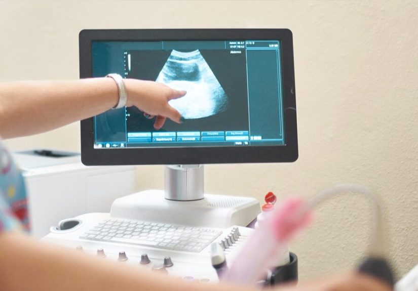
What happens during the ultrasound appointment
Most 16-week ultrasounds are done transabdominally (on your belly). Here’s the typical play-by-play:
- Check-in and setup: You’ll lie back on an exam table.
- Gel time: Warm-ish (sometimes cold) gel is applied to your belly to help the probe transmit sound waves.
- Scanning: The tech moves the transducer over your abdomen to capture images and measurements.
- Freeze frames and measurements: You’ll see the cursor lines and image “snapshots” as they record key views.
- Wrap-up: You wipe off the gel (victory), and your provider reviews results.
Many ultrasound appointments take roughly 30–60 minutes, depending on the type of scan and how cooperative
your baby feels about being photographed that day.
Will it ever be transvaginal at 16 weeks?
Sometimes, yesespecially if your provider needs a closer look at the cervix or placenta location, or if abdominal
views aren’t clear. Transvaginal ultrasound uses a different probe and can provide clearer imaging in certain cases.
If this is planned, the clinic usually tells you beforehand.
What you’ll see at 16 weeks
At 16 weeks, your baby is no longer a “blob with potential” and very much a tiny human-in-progress.
On ultrasound, you may see:
- Head and facial profile: The curve of the skull, nose, and jaw may show up depending on angle.
- Spine and ribs: Bone shows clearly on ultrasound and often looks bright white.
- Arms, legs, hands, and feet: You may catch baby flexing, stretching, or practicing karate.
- Heart motion: You’ll likely see the heartbeat flicker and may see chambers depending on the scan.
- Movement: Many babies are active even before you can consistently feel it.
- Placenta and amniotic fluid: The tech may note placenta position and fluid volume.
- Umbilical cord: Sometimes visible as baby drifts near it.
You may also notice that ultrasound images aren’t “Instagram clear.” They’re more like: “Congratulations, this
abstract art piece is your child.” The sonographer helps translate what you’re seeing, and your provider interprets
it in the context of your pregnancy.
Can you tell the baby’s sex at 16 weeks?
Sometimes. At 16 weeks, external genitalia may be visible, but accuracy depends on fetal position, equipment,
the skill and policies of the clinic, and plain old luck. If baby isn’t in the right position, you may not get a clear
viewand that’s normal. Some people find out at 16 weeks; others have to wait until the anatomy scan.
Also worth noting: if you already had genetic screening (like NIPT), that may provide sex chromosome information
earlier than ultrasoundif you chose to learn it.
What your provider is checking (the “why we’re here” list)
A 16-week ultrasound might include some or all of the following assessments, depending on the goal of the exam:
Growth and gestational age
Measurements may include head size and other standard fetal biometry. These help confirm growth patterns and
support dating if there’s uncertainty.
Fetal heartbeat and general well-being
Ultrasound confirms fetal cardiac activity and can show general movement and tonereassuring signs that things are
progressing.
Placenta location
Placenta position can matter, especially if it’s near the cervix (sometimes called “low-lying placenta” early on).
Many placentas that are low earlier in pregnancy move upward as the uterus grows, so a 16-week scan might be a check-in,
not a final verdict.
Cervical length (when indicated)
In certain pregnancies, your provider may evaluate the cervix to assess risk for preterm birth. This may involve
transvaginal imaging for the clearest measurement.
Amniotic fluid
Fluid volume around baby can be assessed visually or measured, depending on the scan type.
Early anatomy check (sometimes)
While the most comprehensive anatomic survey is typically done a bit later, some centers can evaluate key structures
earlierespecially for high-risk cases or when advanced imaging is available. Even so, it’s common to repeat or complete
anatomy evaluation in the standard 18–22-week window for the fullest picture.
What happens after the ultrasound
After the scan, your provider (OB-GYN, midwife, or maternal-fetal medicine specialist) reviews the images and report.
You might get results:
- Immediately (some offices discuss right away),
- At your next visit, or
- Through your patient portal once the report is finalized.
If they recommend a follow-up scan, don’t panic
Follow-ups are common for simple reasons: baby was in a tricky position, certain views weren’t clear, or your provider
wants to track something (like placenta location) over time. A repeat scan doesn’t automatically mean something is wrong.
Safety and limits: what ultrasound can (and can’t) do
Medical ultrasound uses sound wavesnot radiationto create images. It’s widely used in pregnancy, and major medical
organizations consider it safe when used appropriately.
That said, ultrasound isn’t a crystal ball. It can’t detect every condition, and some findings are unclear until later.
The quality of images can also be affected by fetal position, the amount of amniotic fluid, body tissue, and scarring.
Your provider uses ultrasound results alongside labs, exams, and the full clinical picture.
FAQ: quick answers to common 16-week ultrasound questions
Can I bring someone with me?
Many clinics allow a partner or support person, but policies varyespecially depending on office rules, space, and
infection-control guidelines. Check ahead so you’re not surprised at the door.
Will I get pictures?
Often, yesespecially if it’s a routine-style scan. But if the ultrasound is focused on a medical question, the priority
may be clinical images rather than print-worthy snapshots. You can always ask what the clinic provides.
What if I don’t want to know the sex?
Tell the ultrasound team at the start. They’re used to “team surprise,” and they can often document needed anatomy
without revealing sex-related details.
Is 3D/4D ultrasound included?
Typically, standard medical ultrasounds are 2D. Some clinics may offer 3D/4D views, but they’re not always necessary for
medical evaluation. If you’re considering non-medical “keepsake” ultrasound services, discuss it with your providermedical
ultrasound is recommended when there’s a clinical reason.
Real-life experiences at a 16-week ultrasound (about )
Every pregnancy is different, but people tend to describe 16-week ultrasounds with a similar mix of excitement, nerves,
and “why is my baby doing parkour right now?” Here are some common experiences parents shareso you can feel more prepared
and less like you’re walking into a pop quiz you didn’t study for.
The waiting-room brain spiral
Many people say the hardest part is the few minutes before the scan starts. Your mind can bounce between “I can’t wait to
see the baby!” and “What if they find something?” That emotional whiplash is normal. A helpful strategy some parents use:
pick one grounding thought (like “This appointment is information, and information helps us make good decisions”) and repeat
it when anxiety gets loud.
The gel is… memorable
People almost always comment on the geleither because it’s unexpectedly cold or because it somehow travels farther than
seems physically possible. (If there’s a secret to removing gel from a waistband, science has not found it yet.)
A small towel or wipes are usually provided, but some parents bring an extra tissue just in case.
“Is that the head? Or… a knee?”
Ultrasound images can be confusing at first. Many parents describe a few moments of staring at the screen, smiling politely,
and hoping nobody asks them to identify anything. Then the sonographer points out landmarksskull, spine, limbsand suddenly
it clicks. A common tip: don’t be afraid to say, “Can you show me what I’m looking at?” That’s a normal request.
Baby’s personality makes an early appearance
Some babies stay still long enough to get clear measurements. Others treat the ultrasound like a performance.
Parents often talk about seeing:
- little kicks or stretches that look way stronger than expected,
- hands near the face (classic),
- turning away at the exact moment you want a picture (also classic),
- and the occasional “wave,” which is really just a normal arm movementbut everyone agrees it’s a wave.
Finding out the sex: excitement, uncertainty, or a big “maybe”
If you’re hoping to learn the baby’s sex at 16 weeks, parents describe three common outcomes:
(1) you get a clear view and a confident answer; (2) you get a “probably, but we’ll confirm later”; or (3) baby refuses to
cooperate, and you leave with a new respect for how stubborn someone can be at the size of an avocado.
After the scan: relief, more questions, or both
Many parents feel immediate relief just seeing the heartbeat and movement. Others walk out with follow-up planslike returning
for a repeat scan because views were limited, or scheduling the anatomy scan for the 18–22-week window. The most common theme:
leaving with more connection to the pregnancy than before. For a lot of families, this is the moment the pregnancy starts
feeling real in a new way.
Conclusion
A 16-week ultrasound can be a reassuring checkpoint and an exciting glimpse into your baby’s second-trimester development.
While it may not replace the detailed anatomy scan typically done a bit later, it can answer important questions, confirm
growth, and give you a chance to see movement, heartbeat, and early features. Go in knowing what the scan is for, what it can
(and can’t) confirm, and that “baby wouldn’t hold still” is basically the unofficial motto of prenatal imaging.